Modern Homoeopathy
Monthly E-Newsletter June 2008
Xanthelasma and Homoeopathy
by
Dr. Rajneesh Kumar Sharma, MD (Homoeopathy) & Dr. (Km.) Ruchi Rajput, BHMS
Definition
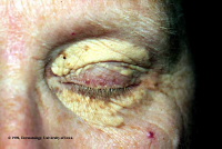
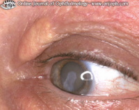
Xanthelasma or Xanthomas are deposits of fat or cholesterol (Sycosis) in the eye lid. These appear as yellowish plaques or nodules in the subcutaneous tissues in the periorbital region. They represent an accumulation of lipid-containing macrophages (Psora) in the dermis. In about 50% of patients lipid levels are normal although in young individuals with this condition there is a higher incidence of hypercholesterolaemia (Sycosis). Possible causes are raised cholesterol (Sycosis), hyperlipidaemia states (Sycosis), diabetes mellitus (Syphilis) and obesity. They cause no harm, but can increase in size producing cosmetic blotch.
Gross and microscopic appearance
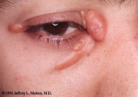
It is of yellow flat plaques over the upper or lower eyelids. In other areas of the body the individual lesion is called xanthoma.
Xanthoma is a tumour composed of lipid-filled histiocytes containing lipid material in the cytoplasm. A prominent manifestation of the hyperlipoproteinaemias is xanthomas in soft tissue, tendinous, subperiosteal and intraosseous locations. Tuberous and tendinous xanthomas produce nodular masses in soft tissue (Sycosis) and tendons that rarely calcify (Sycosis); tendinous xanthomas are common in the fingers, heel, elbow and knee, at which sites they may erode subjacent bone.
Subperiosteal xanthomas are associated with scalloping (Sycosis) of the external cortical surface. Intramedullary deposition of lipids (Psora) leads to lytic defects (Syphilis) and pathologic fractures (Psora/ Syphilis).
Ultrasonography, CT and MR imaging are favoured for the diagnosis of soft tissue xanthomas. The signal intensity of xanthomas on MR imaging varies. The tumours may be of persistent low to intermediate signal intensity on T1-weighted and T2-weighted spin-echo MR images and may show an inhomogeneous signal pattern. Focal areas of high signal intensity may occasionally be encountered on T2-weighted images, however. Some patients reveal a diffuse speckled pattern of signal intensity within a tendon.
Etiology
The possible causes of Xanthelasma formation are-
Pathogenesis
Xanthelasma may follow erythroderma (Psora) and inflammatory skin disorders (Psora) in the presence of normal lipid profiles.
The mechanism that initiates macrophage accumulation (Psora), cholesterol uptake (Psora) and foam-cell formation (Sycosis) in a normolipaemic patient following an inflammatory skin disorder (Psora) is not yet been elucidated a mechanism. This mechanism has been suggested is that increased plasma lipid peroxidation (Psora) (derived from oxidized low-density lipoprotein) may lead to accumulation of cholesterol (Sycosis) in macrophages and formation of foam cells (Sycosis).
Diagnosis
It is easily done since colour and site are characteristic.
Associated Risks
Xanthelasma may be associated with familial hyperlipidaemia (Sycosis). Patients with these lesions therefore frequently also have arcus senilis and xanthomas in other areas of the body. The presence of xanthelasma and corneal arcus indicates a higher risk of developing ischaemic heart disease (Psora/ Sycosis/ Syphilis), but not peripheral vascular disease (Psora).
Secondary hyperlipidaemia (Sycosis) can also be an association, usually caused by underlying uncontrolled diabetes.
Some patients exhibiting xanthelasma have normal lipid levels.
Differential diagnosis and synonymous complaints
Management
The lesions can be removed for cosmetic reasons. Fasting lipid levels are checked, and the patients with hyperlipidaemia should have a formal cardiovascular risk assessment. There is no evidence that lipid lowering treatment has any impact on the appearance of Xanthelasma.
Prognosis
The Xanthelasma itself is harmless. Recurrence after treatment is common. Prognosis is affected by any associated co-morbidity.
Homoeopathic Treatment
Allium-sativum, Aurum metallicum, Baryta muriaticum, Calcarea-fluoricum, Calcarea carbonicum, Chelidonium majus, China officinalis, Chionanthus, Cholestrnum, Chromicum-acidum, Colchicum, Cortisonum, Ferrum-iodatum, Hydrastis, Lecithinum, Medorrhinum, Nux-vomica, Perh-mal, Taraxicum, Thuja occidentalis, Thyreotropinum, Vanadium, Zingiber officinalis
Bibliography
© Dr. Rajneesh Kumar Sharma, MD (Homoeopathy)
Dr. (Km.) Ruchi Rajput, BHMS
Homoeo Cure Research Centre P. Ltd.
NH 74- Moradabad Road
Kashipur (UTTARANCHAL) - INDIA
Ph- 09897618594
|
|